


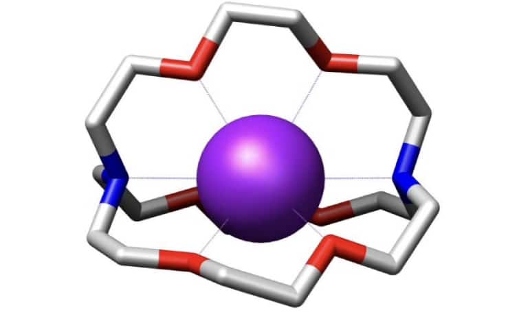
Figure 1. Structure of [2.2.2] Cryptand encapsulating a potassium cation (purple) as determined by X-ray crystallography.
Credit: M stone, CC BY-SA 3.0 via Wikimedia Commons; data from Alberto et al., JACS 2001.
MacroLig® 404 (Cryptand 2.2.2) is a macrocycle widely used in the synthesis of diagnostic radiopharmaceuticals, such as those utilizing Fluorine-18 (18F). Building on decades of Molecular Recognition Technology® (MRT™) and large-scale macrocycle manufacturing expertise, IBC Advanced Technologies develops advanced, custom macrocycles designed specifically for therapeutic radioligand applications. IBC is leveraging its proprietary macrocyclic chemistry and molecular design to deliver custom chelators optimized to securely bind high-demand therapeutic isotopes, including Radium-223 and Radium-224 (223Ra/224Ra), Actinium-225 (225Ac), and Lead-212 (212Pb). Beyond these isotopes, IBC’s macrocyclic design platform supports a wide range of therapeutic isotopes across alpha, beta, and Auger-emitting radiopharmaceuticals, enabling custom chelators tailored to the specific coordination chemistry of each isotope.
Custom Macrocycles: The Hidden Magic Bullets Driving Precision Radioligand Therapy
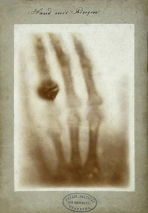
The history of radiation collided with medical history in 1895, when Wilhelm Roentgen found that a previously undiscovered “ray” was being emitted from his laboratory’s cathode-ray tube when high voltage was applied. He noticed that this new ray was capable of passing through most solid objects, but not bone or metals. The first “roentgen photograph”, showing the bones of his wife’s hand, was produced shortly afterwards. Soon after, capturing the radioactivity uranium and radium gave off with photography earned Henri Becquerel a shared Nobel Prize with Maria Skłodowska-Curie and Pierre Curie in 1903. These parallel discoveries allowed the proliferation of radioactivity in medicine [1].
The complications of using radiation were noticed early on. Pierre Curie noted in his Nobel speech that, “If one leaves a small glass ampulla with several centigrams of radium salt in one’s pocket for a few hours, one will feel absolutely nothing. But in 15 days afterwards redness will appear on the epidermis, and then a sore, which will be very difficult to heal. A more prolonged action could lead to paralysis and death.” [2]. Perhaps it was the clear damage to the skin that radiation was capable of causing that gave the idea for cancer treatments so quickly. Emil Grubbe in the United States and Victor Despeignes in France are both recorded as having begun using X-rays for medical imaging and cancer treatment in 1896, only months after Wilhelm Roentgen publicized his findings [3]. Grubbe lived out a short retirement, with the losses of his right hand, nose, upper lip, and most of the right side his face to cancer caused by excessive exposure to radiation. During his career he taught 7,000 other doctors how to use X-rays and pioneered the use of radiation to decrease and remove cancerous tumors [3].
Using radioactivity for cancer treatment has always been a practice of balancing on a razor’s edge. Radiation is inherently dangerous to living cells of all kinds. It does its damage at the most basic, minute level, by breaking bonds of DNA. Because radiation exists all around us, and always has, cells are able to heal from low levels of radiation. However, given enough time, even low levels of radiation damage remain unhealed, or heal incorrectly, and that damage at the cellular level will eventually turn into cancer [4]. The fundamental truth of toxicology remains: the dose makes the poison. While uncontrolled radiation has long been known to cause cancer, it can now be used against the disease in carefully structured and localized dosages to destroy malignant cells while leaving healthy surrounding tissue unharmed.
Moving from a Hail of Bullets to a Sniper Shot
Cancerous cells are living cells too, and just as vulnerable to broken DNA as any other. When targeted correctly, radiation can be used strategically against cancer cells while sparing the surrounding tissue from most of the danger. Historically, however, radiation therapy was a blunt instrument, a literal shower of DNA-destroying particles hitting both healthy and diseased tissue alike. As time has gone on, the goal has been to move from the hail of DNA-destroying bullets, to an impossibly precise sniper shot, directly to the cancerous cells, completely bypassing all other cells in the body.
At the dawn of our modern age of medicine, Paul Ehrlich, a German physician and researcher, reasoned that if a compound could be made that selectively targeted a disease-causing organism, then a toxin for that organism could be delivered along with the agent of selectivity. Hence, a “magic bullet” would be created that killed only the organism targeted [5]. His reasoning has proved correct and “magic bullets” have been found for many diseases. For certain types of cancer, radioligand therapy is the closest humankind has yet reached into finding a magic bullet for cancer.
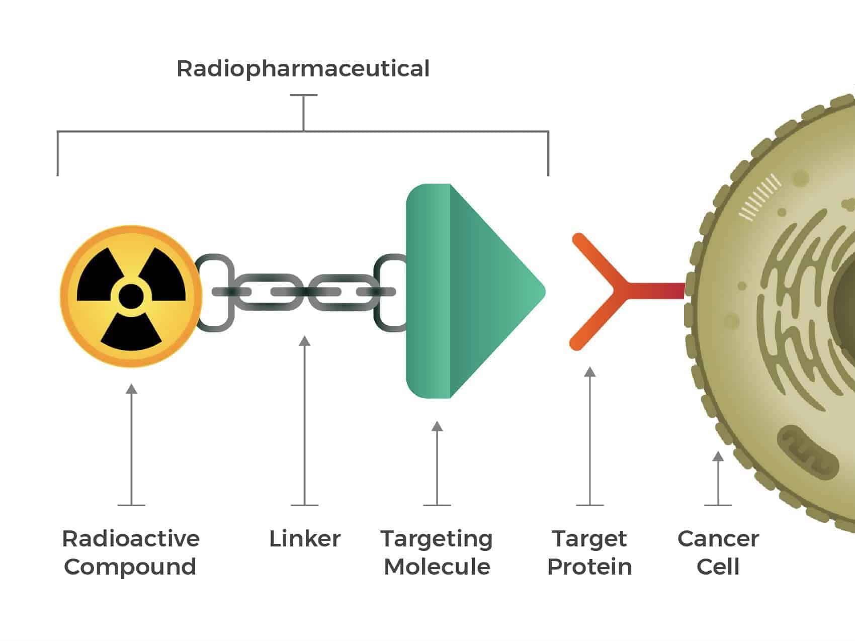
The Three Essential Components of Radioligand Therapy
Radioligand therapy is a cancer therapy that targets and delivers a lethal dose of radiation to a tumor. The injectable radiopharmaceutical consists of three key components. First, the radioactive compound which is made up of a radioisotope that emits alpha or beta particles and a chelator that strongly binds the radioisotope. Second, the targeting molecule or ligand which recognizes and binds to cancer cells. Third, the linker which connects the radioactive compound to the targeting molecule. All three components are essential to deliver the destructive radiation directly to cancer cells. For example, if the chelator does not bind strongly to the radioisotope, then deadly radiation will be delivered to other, healthy cells. To minimize these off-target effects, appropriate chelators must be selected. Numerous chelators suitable for small radioisotopes have been established to date, but chelators that work well for large radioisotopes are significantly less common.
Why Modern Radiopharmaceuticals Depend on Macrocyclic Chemistry
There is no universal chelator. Just as ligands and radioactive isotopes are chosen from many options for each type of cancer, the chelator that will link them needs to be just right for the specifics of the case. In some cases, commonly stocked and cheap chelating options like EDTA and DTPA are usable, but inferior. They are open-chain linear compounds that are loose and much more likely to lose grip of either the radioactive compound or the targeting molecule, or both.
Superior in use, but harder to manufacture, are macrocycles: large, cyclic molecules that form a closed ring. Their large size and closed shape link the radioactive isotope and targeting ligand very securely. DOTA and Macropa are two macrocyclic compounds that can complex some radioactive metals very well. Two derivatives of DOTA are currently used for targeted radioligand therapy to bind the beta-emitting radionuclide lutetium-177: Lutathera and Pluvicto.
Now, much research is devoted to developing chelators for alpha-emitting radionuclides such as actinium-225 and lead-212. Ensuring high thermodynamic stability and rapid kinetics for complexation is critical for their use in radioligand therapy. The synthesis of these macrocyclic compounds is complex, and few manufacturers can produce these chelators with high consistency at scale.
RadSep and IBC: Manufacturing the Next Generation of Precision Medicine
IBC Advanced Technologies, Inc. (IBC) specializes in developing and manufacturing highly selective macrocyclic chelators at scale through its subsidiary company, RadSep, Inc. Chelators for actinium, lead, bismuth and radium are being produced, including a class of compounds that can be better than Macropa for binding radium in serum. RadSep’s products are not only precisely manufactured in any quantity but can also be customized, and this ability greatly expands the potential of radioligand therapy. As new ligands and radioisotopes are explored for their usability in medicine, chelators that are suitable for each pair must be found, and RadSep uniquely combines decades of experience in this field with an industry-leading reputation for precision and reliability. RadSep is able to ensure the selectivity and effectiveness of commonly used chelators while expanding the potential library of radioligand therapy by providing custom synthesis for structures that are not in any catalog.
Ready to advance your therapeutic pipeline? Leverage decades of Molecular Recognition Technology® (MRT™) to secure your targeted alpha or beta therapy. Contact the RadSep Custom Synthesis Team to discuss a custom macrocycle tailored to your target isotope, or explore our established diagnostic line by viewing the IBC MacroLig® Product Catalog.
Sources:
[1] Reed, A.B. (2011). The history of radiation use in medicine. Journal of Vascular Surgery, 53(1), pp.3S-5S. doi:10.1016/j.jvs.2010.07.024. Available online at: https://www.sciencedirect.com/science/article/pii/S0741521410017271
[2] Nobel lectures, physics 1901–1921. Elsevier Publishing Company, Amsterdam (1967). Available online at: https://ia801405.us.archive.org/15/items/in.ernet.dli.2015.147948/2015.147948.Nobel-Lectures-Physics-1901-1921_text.pdf
[3] Pioneer in X-Ray Therapy. (1957). Science, 125(3236), 18–19. Available online at: http://www.jstor.org/stable/1752791
[4] CDC (2024). About Health Effects of Radiation. Radiation and Your Health. Available online at: https://www.cdc.gov/radiation-health/about/health-effects-of-radiation.html [Accessed 9 Sept. 2026].
[5] Science History Institute (n.d.). Paul Ehrlich. Science History Institute. Available at: https://www.sciencehistory.org/education/scientific-biographies/paul-ehrlich/ [Accessed 9 Sept. 2026].